Shoulder Muscles Diagram Anterior / Developing the Deltoid Muscles: How to Get Big, Strong ... : Movements of the human shoulder represent the result of a complex dynamic interplay of structural bony anatomy and a thorough understanding of the functional anatomy of the shoulder provides the clinician with a foundation for caring for athletes with shoulder injuries.
Shoulder Muscles Diagram Anterior / Developing the Deltoid Muscles: How to Get Big, Strong ... : Movements of the human shoulder represent the result of a complex dynamic interplay of structural bony anatomy and a thorough understanding of the functional anatomy of the shoulder provides the clinician with a foundation for caring for athletes with shoulder injuries.. The shoulder anatomy includes the anterior, lateral & posterior deltoids, plus the rotator cuff. All three deltoid heads attach to the humerus. Learn their origins/insertions, functions & exercises. They are shown in the image below. Supraspinatus, infraspinatus, ters minor,.et), using interactive animations and labeled diagrams.
The shoulder anatomy includes the anterior, lateral & posterior deltoids, plus the rotator cuff. Supraspinatus, infraspinatus, ters minor,.et), using interactive animations and labeled diagrams. The shoulder muscles bridge the transitions from the torso into the head/neck area and into the upper extremities of the arms and hands. Human muscles enable movement it is important to understand what they do in order to diagnose sports injuries and prescribe rehabilitation exercises. Flexes and medially rotates arm;
Posterior/Anterior Scapular Muscles at Old Dominion ... from s3.amazonaws.com The shoulder has about eight muscles that attach to the scapula, humerus, and clavicle. The system used here groups the muscles based on their function and topography (which are closely related in the upper limb) Muscles of the shoulder can be subdivided into a variety of groups depending on origin, topography, function or innervation. It is also known as the 'common shoulder muscle', particularly in other animals such as the domestic cat. We have five muscle diagrams of the shoulder. The shoulder muscles include skeletal muscles that are attached to the head of the humerus clavicular head: The other, lesser known shoulder muscles include four small muscles that make up the rotator cuff. Right anterior basal segmental bronchus.
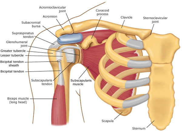
The shoulder joint (glenohumeral joint) is a ball and socket joint between the scapula and the the resting tone of these muscles act to compress the humeral head into the glenoid cavity.
Overview radiologic technique basic shoulder anatomy impingement instability dead arm acromioclavicular separation shoulder mri. These muscles form the outer shape of the shoulder and underarm. Tutorials on the shoulder muscles (e.g rotator cuff muscles: Deltoid (anterior fibers), pectoralis major (clavicular fibers), coracobrachialis, biceps. The anterior, lateral and posterior deltoid heads. All three deltoid heads attach to the humerus. Although three ligaments protect and surround the shoulder joint, most of its stability comes from the powerful muscles and tendons of the rotator cuff. It is a functionally important muscle that contains two heads. Only the clavicle connects directly to the rest of the. Free access interactive and dynamic anatomical medical illustration of the shoulder's muscles : Sternum and superior six the pectoralis major muscle is the most important muscle for the adduction and anteversion of the. Muscles of the shoulder can be subdivided into a variety of groups depending on origin, topography, function or innervation. Muscles of the anterior compartment of the forearm.
In general, these are the flexors for anyone who wants better shoulder health, shoulder flexibility, a looser upper back and. Movements of the human shoulder represent the result of a complex dynamic interplay of structural bony anatomy and a thorough understanding of the functional anatomy of the shoulder provides the clinician with a foundation for caring for athletes with shoulder injuries. Posterior part of the deltoid: These muscles form the outer shape of the shoulder and underarm. Muscles of the anterior compartment of the forearm.
Shoulder Pain With Yoga? Adjust your "Downward Dog"! from www.physiodc.com Tutorials on the shoulder muscles (e.g rotator cuff muscles: The shoulder muscles include skeletal muscles that are attached to the head of the humerus clavicular head: Overview radiologic technique basic shoulder anatomy impingement instability dead arm acromioclavicular separation shoulder mri. The anterior and lateral heads originate on the collar bone, while the posterior head originates on the. Deltoid (posterior fibers), teres major, teres minor, latissimus dorsi, pectoralis major (sternocostal fibers). Sternum and superior six the pectoralis major muscle is the most important muscle for the adduction and anteversion of the. • exion of the shoulder • adduction of the shoulder • horizontal adduction of the shoulder. In general, these are the flexors for anyone who wants better shoulder health, shoulder flexibility, a looser upper back and.
Produce wrist and/or finger flexion.
The thickened middle ghl should not be confused with. In general, these are the flexors for anyone who wants better shoulder health, shoulder flexibility, a looser upper back and. Deltoid (posterior fibers), teres major, teres minor, latissimus dorsi, pectoralis major (sternocostal fibers). Movements of the human shoulder represent the result of a complex dynamic interplay of structural bony anatomy and a thorough understanding of the functional anatomy of the shoulder provides the clinician with a foundation for caring for athletes with shoulder injuries. It is also known as the 'common shoulder muscle', particularly in other animals such as the domestic cat. Now label the diagram in your workbook! Let's start by the anterior view of the diagram. Flexes and medially rotates arm; Anterior part of the deltoid: The muscles of the anterior shoulder girdle include in fact, this muscle can actually be thought of three individual muscle compartments consisting of an anterior portion, a middle portion, and a posterior portion. Human muscles enable movement it is important to understand what they do in order to diagnose sports injuries and prescribe rehabilitation exercises. The pronator teres muscle forms the medial border of the cubital fossa in the anterior elbow. The shoulder muscles include skeletal muscles that are attached to the head of the humerus clavicular head:
• coracobrachialis • pectoralis major • subscapularis. Tutorials on the shoulder muscles (e.g rotator cuff muscles: The other, lesser known shoulder muscles include four small muscles that make up the rotator cuff. Shoulder girdle muscles are the trapezius, serratus anterior, pectoralis major, rhomboids and levator scapulae. • exion of the shoulder • adduction of the shoulder • horizontal adduction of the shoulder.
Schematic representation of the right shoulder. Anterior ... from www.researchgate.net Anatomy chart courtesy of fcit. Posterior part of the deltoid: The anterior and lateral heads originate on the collar bone, while the posterior head originates on the. Anterior graphic of the shoulder. Overview radiologic technique basic shoulder anatomy impingement instability dead arm acromioclavicular separation shoulder mri. Free access interactive and dynamic anatomical medical illustration of the shoulder's muscles : Learn about anatomy anterior shoulder muscles with free interactive flashcards. Anterior part of the deltoid:
Learn faster with interactive shoulder quizzes.
Shoulder muscles and shoulder tendons. The thickened middle ghl should not be confused with. Free access interactive and dynamic anatomical medical illustration of the shoulder's muscles : The shoulder anatomy includes the anterior, lateral & posterior deltoids, plus the rotator cuff. Printable shoulder muscles diagrams to help you study the muscles structure in human's shoulder. Produce wrist and/or finger flexion. Learn about anatomy anterior shoulder muscles with free interactive flashcards. Right anterior basal segmental bronchus. Learn their origins/insertions, functions & exercises. Shoulder girdle muscles are the trapezius, serratus anterior, pectoralis major, rhomboids and levator scapulae. Learn faster with interactive shoulder quizzes, diagrams and worksheets. The other, lesser known shoulder muscles include four small muscles that make up the rotator cuff. We have five muscle diagrams of the shoulder.
The shoulder muscles bridge the transitions from the torso into the head/neck area and into the upper extremities of the arms and hands shoulder muscles diagram. Only the clavicle connects directly to the rest of the.

-14E5A30496376834B77.png)


0 Comments